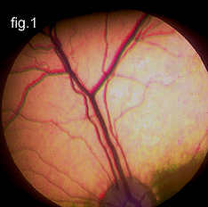

Fig. 1. The normal retina. Note the many prominent blood vessels.
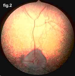
Fig. 2. A mid-stage PRA retina. Notice how the vascularity has been markedly reduced.
Progressive retinal atrophy, or PRA as it is frequently termed, is a long recognized, hereditary, blinding disorder. It is inherited as a simple autosomal recessive in most breeds. The first modern description of this problem was in Gordon Setters in Europe, in 1911, but since then PRA has been recognized in most purebred dogs. Millichamp et al. In 1988, described PRA in Tibetan Terriers. Also in 1988, it was found that PRA in Cockers, Poodles and Labradors was the result of a mutation at the same gene locus in all these breeds.
PRA is a disease of the retina. This tissue, located inside the back of the eye, contains specialized cells called photoreceptors that absorb the light focused on them by the eye’s lens, and converts that light, through a series of chemical reactions into electrical nerve signals. The nerve signals from the retina are passed by the optic nerve to the brain where they are perceived as vision. The retinal photoreceptors are specialized into rods, for vision in dim light (night vision), and cones for vision in bright light (day and color vision). PRA usually affects the rods initially, and then cones in later stages of the disease. In human families, the diseases equivalent to PRA (in dogs) are termed retinitis pigmentosa.
In all canine breeds PRA has certain common features. Early in the disease, affected dogs are nightblind, lacking the ability to adjust their vision to dim light; later their daytime vision also fails. As their vision deteriorates, affected dogs will adapt to their handicap as long as their environment remains constant, and they are not faced with situations requiring excellent vision. At the same time the pupils of their eyes become increasingly dilated, in a vain attempt to gather more light, causing a noticeable "shine" to their eyes; and the lens of their eyes may become cloudy, or opaque, resulting in a cataract.
The big difference in PRA among breeds is in the age of onset and the rate of progression of the disease. Certain breeds, notably including the Collie, the Irish Setter, the Norwegian Elkhound and the Miniature Schnauzer, have early onset forms. In these breeds the disease results from abnormal or arrested development of the photoreceptors—the visual cells in their retina, and affects pups very early in life. In other breeds, including the Miniature Poodle, the English and American Cocker Spaniel, and the Labrador Retriever, and many other breeds, including the Tibetan Terrier, Tibetan Spaniel, and Lhasa Apso, PRA is much later in onset. Affected dogs in these breeds appear normal when young, but develop PRA as adults.
Diagnosis of PRA is normally
made by ophthalmoscopic examination.
This is undertaken using an instrument called an indirect
ophthalmoscope,
and requires dilatation of the dog’s pupil by application of
eyedrops.
Broadly speaking, all forms of PRA have the same sequence of
ophthalmoscopic
changes: increased reflectivity (shininess) of the fundus (the inside
of
the back of the eye, overlain by the retina); reduction in the diameter
and branching pattern of the retina’s blood vessels; and
shrinking of the
optic nerve head (the nerve connecting the retina to the brain). These
changes occur in all forms of PRA, but at different times in the
different
breed-specific forms. Usually by the time the affected dog has these
changes
there is already significant evidence of loss of vision.
 |  |
Fig. 1. The normal retina. Note the many prominent blood vessels. |
Fig. 2. A mid-stage PRA retina. Notice how the vascularity has been markedly reduced. |
Confirmation of the diagnosis can be undertaken by electroretinography. This is an electrical measurement of retinal function somewhat similar to an electrocardiographic test of heart function, but with two differences: the electroretinogram (ERG) can only be recorded as a response to a flash of light (ie: it is not a free running signal like the EKG); and accurate recording of the ERG requires that the dog be anesthetized. In all dogs showing clinical evidence of PRA, the ERG is severely diminished or extinguished. The ERG can also be used for early diagnosis of specific forms of PRA, that is to detect PRA-affected dogs before they demonstrate clinical evidence of disease. This requires very carefully controlled ERG recording conditions, and a well defined understanding of the age of onset and rate of change of ERG dysfunction in the specific form of PRA under consideration.